

 Current
Location:
Animal Disease Diagnostics: Assays, Instruments and Workflow | HENG MEI Intelligent Manufacturing
Article Source: Hengmei Technology Release time:2026-11-16 16:51:42
An animal disease diagnosis is never produced by an instrument alone. It is produced by a chain: a sample that represents the animal, a reagent that recognises the target, an instrument that measures the reaction, and a rule that turns the measurement into a reportable result. Weakness at any step invalidates the others. This article explains each link in that chain, defines the terms that appear on specification sheets, and shows what the instrument does — and does not do — at each stage.
HENG MEI Intelligent Manufacturing builds the instrument end of that chain: microplate readers for enzyme immunoassay, readers for colloidal gold strips, and real-time PCR instruments. Understanding the whole chain is what allows a buyer to interpret, for example, why a strip reader and a plate reader can both be described as immunoassay instruments yet produce completely different kinds of result.
Antigen and antibody: the two targets that define the method
Almost every veterinary diagnostic test detects one of two things. An antigen is a component of the pathogen itself — a viral capsid protein, a bacterial surface polysaccharide, a toxin. An antibody is the host's own protein produced in response to infection or vaccination, of the immunoglobulin G or M class in most assays. The distinction is the first question to settle, because it determines what the result means.
Detecting antigen answers "is the pathogen present now". Detecting antibody answers "has this animal been exposed or vaccinated". A positive antibody result in an unvaccinated herd indicates exposure; the same result in a vaccinated herd may indicate nothing more than successful vaccination, unless the assay is designed to distinguish marker proteins that vaccine strains lack. This is why tests for pathogens such as pseudorabies are sometimes configured to detect a specific glycoprotein, and why the same species may need two different kits for two different questions.
Why the assay, not the instrument, determines specificity
The specificity of a test — its ability to avoid reacting to the wrong target — comes from the antibody or antigen used as the capture reagent. The instrument measures colour or fluorescence; it does not decide what reacted. A plate reader with excellent optics reading a poorly characterised kit will report a precise number for the wrong measurement. Buyers therefore evaluate a kit and an instrument as a pair, and a demonstration should always use the kits actually intended for field use.
Immunoassay: the same principle, two formats
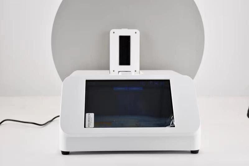
Microplate ELISA
In a sandwich ELISA, capture antibody bound to the well surface binds target antigen from the sample; a detection antibody conjugated to an enzyme binds the captured antigen; a substrate produces colour in proportion to the amount of enzyme present. In an indirect ELISA for antibody detection, the well is coated with antigen and the sample antibody, if present, is detected by an enzyme-labelled anti-species conjugate.
The measured quantity is optical density (OD), also written as absorbance, read at a defined wavelength — commonly 450 nm for horseradish peroxidase systems with a TMB substrate, and 405, 492 or 630 nm in other configurations. OD is not a concentration. Converting OD into a result requires a calibration curve, a single-point calibrator, or a cut-off formula supplied by the kit manufacturer.
Readers measure each well through an optical path. Eight-channel instruments read eight wells simultaneously and step across the plate, so a 96-well plate requires twelve measurement steps; an independent reference channel corrects for lamp drift and well-to-well optical variation. Wavelength selection matters: reading at the substrate's absorbance maximum maximises signal, while a reference wavelength reduces optical interference from the plate plastic and from bubbles.
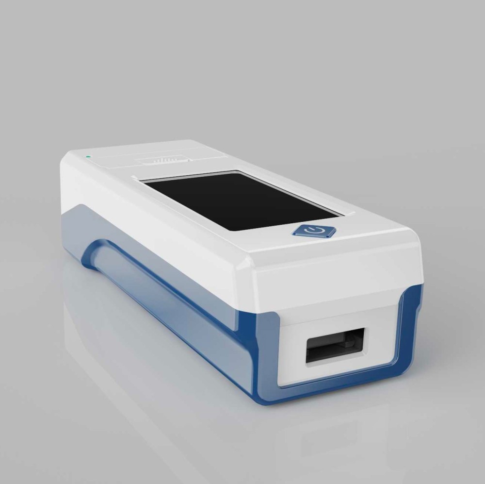
Colloidal gold lateral flow
A lateral flow strip is chromatography rather than a plate assay. Gold-conjugated antibody dried on a conjugate pad is rehydrated by the sample and flows along a membrane. At the test line, immobilised antibody captures the complex if antigen is present, producing a visible red line; at the control line, a second reaction confirms that the strip ran correctly. Results are available in minutes and require no washing, no incubation control and no plate handling.
Reading by eye is the weakest link in that chain. Line intensity varies with lighting, with the operator, and with the degree of gold aggregation, and a faint line is exactly the case where a decision matters most. A strip reader removes that variability by scanning the strip optically, identifying the control and test lines automatically, and reporting a computed index against a stored cut-off. Readers do not make a weak strip stronger; they make the judgement consistent and recordable.
Nucleic acid amplification: detecting the pathogen genome
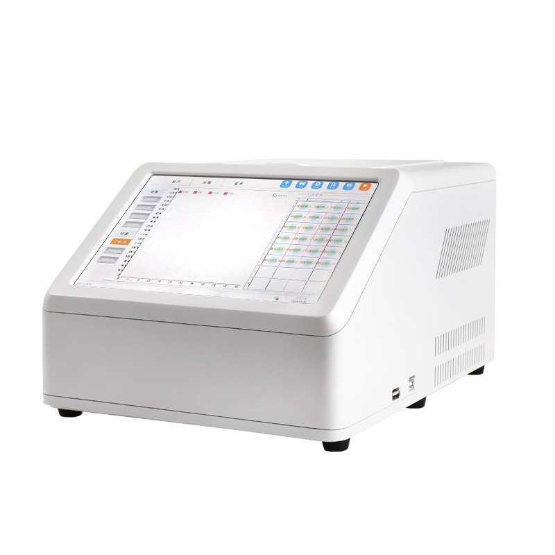
PCR copies a target sequence exponentially through repeated cycles of denaturation, primer annealing and extension. In real-time PCR, a fluorescent reporter signal is measured at every cycle, so amplification is observed as it happens rather than by running a gel afterwards. Three terms dominate the vocabulary.
- Ct value — the cycle number at which fluorescence crosses a threshold. A low Ct means the target was abundant at the start; a high Ct means it was scarce. Ct is therefore a semi-quantitative measure of pathogen load, meaningful only within the linear range of that specific assay.
- Fluorescence channel — the wavelength band in which a particular dye is detected. Two channels allow two targets, or a target plus an internal control, in the same tube.
- Internal control — a template added to each reaction to prove that amplification was not inhibited by the sample matrix. Without it, a negative result cannot be distinguished from a failed reaction.
Instrument specifications that matter for PCR are thermal rather than optical: ramp rate determines run time, control accuracy and well-to-well uniformity determine whether replicates agree, and the number of wells determines daily capacity. A 16-well instrument running 40 cycles with a 3.5 °C/s ramp completes a typical protocol in well under two hours, which is what makes same-day confirmation practical.
The sample workflow, step by step
Every result inherits the quality of the sample. In China, NY/T 541-2016 sets out requirements for the collection, preservation and transport of veterinary diagnostic samples, and it is worth reading before specifying an instrument, because it defines the conditions the instrument's input must satisfy.
Step 1 — Collection that represents the animal
Serum is the standard matrix for antibody testing, and blood must be allowed to clot and be separated promptly; haemolysis interferes with colorimetric reading and, in severe cases, makes an ELISA result uninterpretable. Swabs for antigen or nucleic acid testing must reach the target tissue — nasal, tracheal, rectal or cloacal depending on the disease — and must be placed immediately into the transport medium specified by the kit. Tissue samples for PCR should be chilled rapidly; nucleic acid degrades within hours at summer ambient temperatures.
Step 2 — Transport under a defined cold chain
Sample integrity is a transport property as much as a laboratory property. Insulated boxes with ice packs, a documented dispatch time, and a receiving record convert an informal handover into a traceable chain. Where samples routinely arrive more than 24 hours after collection without cooling, the most valuable purchase is not a more sensitive detector but a transport protocol.
Step 3 — Preparation appropriate to the method
ELISA preparation is comparatively simple: dilution of serum into the kit buffer, and for tissue samples a homogenisation step followed by clarification. Colloidal gold preparation is simpler still. PCR preparation is the demanding stage — nucleic acid extraction, quantification, and set-up in a clean area physically separated from amplification — and it is the stage most likely to be underestimated when a laboratory adds molecular testing for the first time.
Step 4 — Measurement
Measurement is where the instrument enters. A plate reader reports OD per well against a blank and a reference wavelength; a strip reader reports line intensities and a computed result; a PCR instrument reports fluorescence per cycle and derives Ct. In all three cases the raw measurement is the instrument's contribution, and the interpretation belongs to the assay and to the programme's decision rule.
Step 5 — Interpretation against a stated rule
A cut-off converts a number into a classification. Common approaches are a fixed OD threshold, a percentage of a positive control, a sample-to-positive ratio, or an interpolated concentration from a four-parameter curve. Whichever is used, the rule must be written down, applied identically to every batch, and accompanied by control results that demonstrate the run was valid. Two laboratories using different cut-off rules on the same sample can legitimately report different classifications.
Method and target: a reference matrix
| Method | Typical target | Output type | Time to result | Site requirement |
|---|---|---|---|---|
| Colloidal gold lateral flow | Pathogen antigen, or antibody in some formats | Qualitative, with computed index on a reader | 10–20 minutes | Pen side, no laboratory |
| ELISA, 96-well plate | Antibody (serology) or antigen | Quantitative OD, titre or S/P ratio | 2–4 hours including incubation | Bench, washer, incubator, pipettes |
| Real-time PCR | Pathogen nucleic acid | Quantitative Ct, positive or negative | 2–3 hours including extraction | Separated clean area, extraction, cold chain |
| Bacterial culture and identification | Viable bacteria | Isolate, species, antimicrobial susceptibility | 24–72 hours | Microbiology laboratory |
The matrix explains why programmes combine methods rather than replacing one with another. Culture remains necessary for antimicrobial susceptibility, which no immunoassay or PCR can provide. PCR detects nucleic acid whether or not the organism is viable, which is an advantage for fastidious pathogens and a limitation when the question is whether an animal is still infectious.
Analytical terms that are routinely confused
| Term | Definition | Why it matters in procurement |
|---|---|---|
| Analytical sensitivity | Smallest amount of target the assay can detect reliably | Set by the kit and the method, not by the reader |
| Diagnostic sensitivity | Proportion of truly infected animals that test positive | Depends on sample timing and matrix as much as on the assay |
| Diagnostic specificity | Proportion of truly uninfected animals that test negative | Drives false-positive rate and confirmation workload |
| Repeatability | Agreement between repeats under identical conditions | The instrument contribution; quoted as % or OD |
| Reproducibility | Agreement between laboratories or operators | Requires standard operating procedures, not just instruments |
| Limit of detection | Lowest concentration distinguished from blank with stated confidence | Must be quoted for a specific matrix and protocol |
A reader specification such as repeatability ≤0.2% describes instrument precision, not diagnostic accuracy. Buyers who conflate the two end up expecting a plate reader to compensate for a poor kit — which it cannot do.
What a well-run testing chain looks like
Four properties distinguish a programme that produces trustworthy results from one that produces numbers.
- Every batch has controls. Positive and negative controls, and where the kit provides them, calibrators. A batch without valid controls is not a result, whatever the instrument reports.
- Quality control rules are applied, not just recorded. Westgard multi-rule evaluation and quality control charts turn a series of control values into a decision about whether the run is in control.
- Raw data is retained. Auditors and reference laboratories ask for the original OD values, not only the final classification. Instruments that store plate-level records and export them satisfy that requirement without transcription.
- Records carry identity. Sample number, species, premises, collection date, assay name, kit lot and operator must travel with the result. A result that cannot be traced to a sample and a lot cannot support a regulatory decision.
Where HENG MEI instruments sit in this chain
HENG MEI Intelligent Manufacturing supplies the measurement stage in each of the three main formats. The HM-DWYB portable colloidal gold system covers on-site qualitative testing with automatic control and test line recognition, 200,000-result storage and Wi-Fi upload. The HM-Y96A and HM-Y96B microplate readers cover 96-well ELISA in qualitative cut-off and quantitative regression modes, with the Y96B adding a 330–1100 nm range, ten filter positions, eight optical channels plus reference, and a 10.4-inch control surface. The HM-P16 covers 16-well two-channel real-time PCR for confirmation and typing work.
All three produce records that can be exported and archived, which is the property that matters when a programme is later audited or when a result has to be defended to a buyer of livestock. Buyers who want to compare these instruments with international brands can read our brand comparison review; buyers who already know which method they need can go directly to model selection.
Contact: Kyra, Sales Contact, Kyra@hmdzkj.cn, +86 13792642016 (Tel / WhatsApp / WeChat).
Address of this article:https://www.kjhm.net/news/animal-disease-diagnostics-assays-instruments-workflow.html
 Related News
-
24 / 2025-12
HM-Y100 Portable Near-Infrared Rapeseed Analyzer Product Knowledge Graph White Paper
Learn more
-
12 / 2024-11
Unlock the Secrets of Photosynthesis with Portable Photosynthesis Meter.
Learn more
-
21 / 2024-11
Multifunctional pesticide residue detectors can adapt to diverse detection needs.
Learn more
-
13 / 2026-11
HENG MEI Intelligent Manufacturing Choosing an Automatic Microplate Washer
Learn more