
 Current
Location:
Current
Location: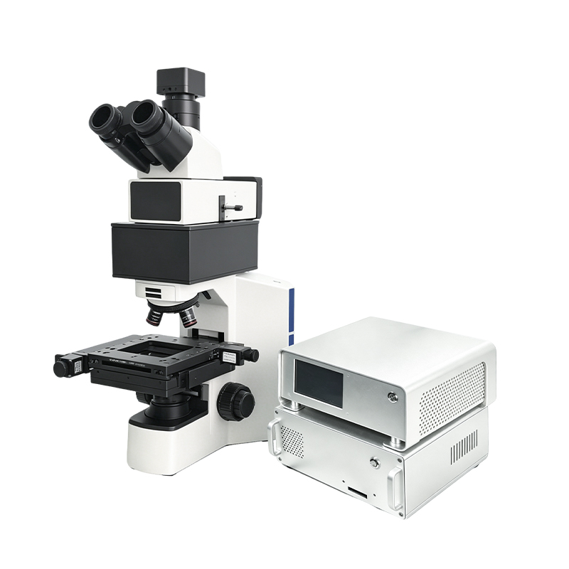
 +86 17853698681
 WhatsApp
 Product Details
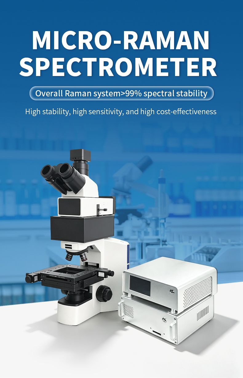


Product Introduction
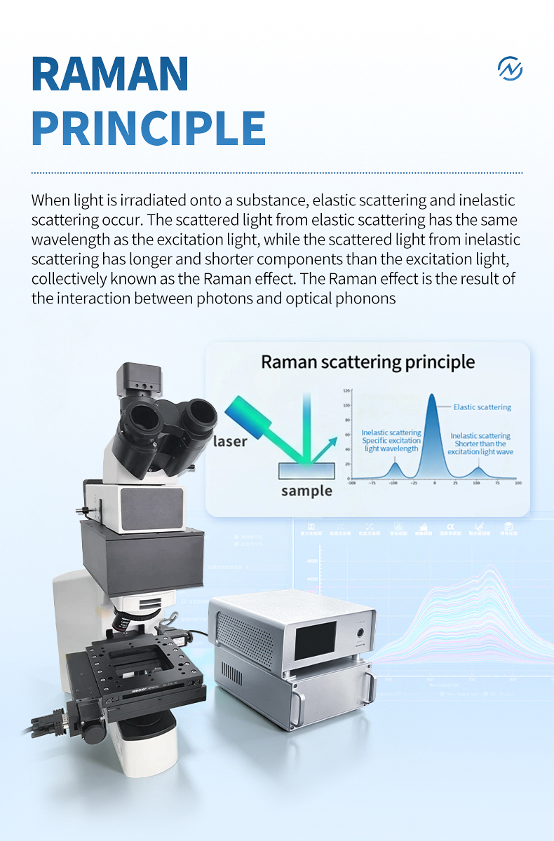
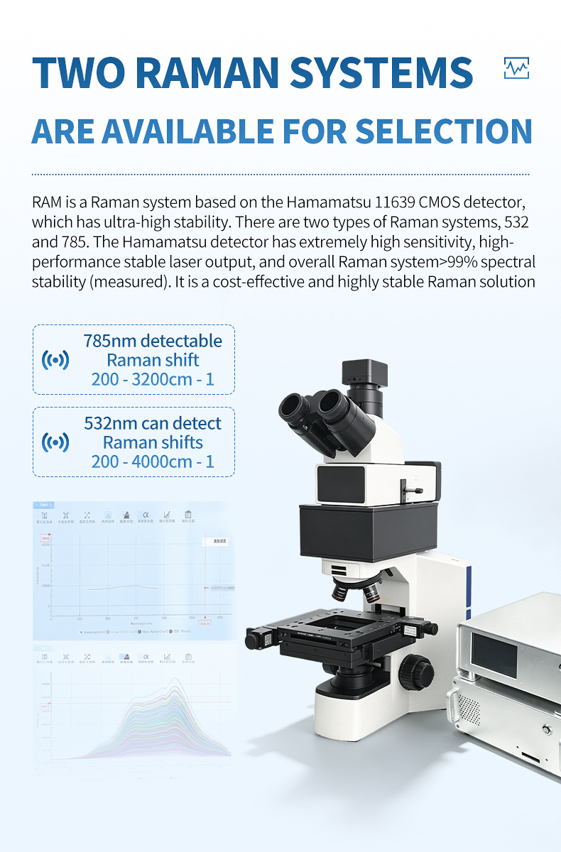
The MR-785B is HM Instruments' flagship micro Raman spectrometer, representing the convergence of near-infrared fluorescence suppression and fully automated spatial mapping. Designed for the most demanding pharmaceutical, biomedical, and advanced materials laboratories, this system combines 785 nm excitation — which virtually eliminates autofluorescence from organic and biological samples — with a precision motorized stage capable of unattended large-area chemical imaging.
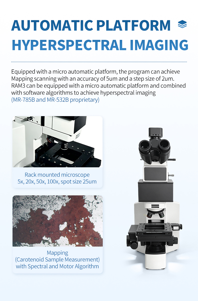
The motorized X-Y platform achieves 5 μm positioning accuracy with 2 μm minimum step size across a 110 mm × 75 mm travel range, while integrated autofocus tracking compensates for surface irregularities during extended mapping sequences. The 1200 gl/mm volume phase holographic grating blazed at 800 nm, coupled with the Hamamatsu 11639 CMOS detector (2048 pixels, >99.8% linearity), delivers a wavenumber range of 200–3200 cm⁻¹ with 7 cm⁻¹ resolution — ideal for distinguishing pharmaceutical polymorphs, mapping tissue biochemistry, and characterizing complex organic mixtures.
Whether analyzing drug tablet homogeneity, mapping microplastic contamination on filter membranes, or conducting high-throughput screening of fluorescence-prone geological samples, the MR-785B provides the automation, sensitivity, and spectral quality required by leading-edge research and quality control facilities. See our Raman industry applications whitepaper for detailed use cases across sectors.
Applications
- Automated polymorph distribution mapping across pharmaceutical tablet surfaces and cross-sections
- High-throughput tissue section imaging for cancer margin assessment and pathological research
- Large-area microplastic particle scanning on environmental filter membranes with automated classification
- Drug coating uniformity and layer thickness mapping for quality control in pharmaceutical manufacturing
- Forensic trace evidence mapping with automated particle detection and spectral library matching
- Geological fluid inclusion and mineral phase mapping in fluorescence-prone host minerals
- Polymer blend morphology and additive distribution analysis across industrial sample areas
- Cultural heritage artifact analysis — pigment and binder mapping on fluorescence-rich historical surfaces
Functional Performance and Features
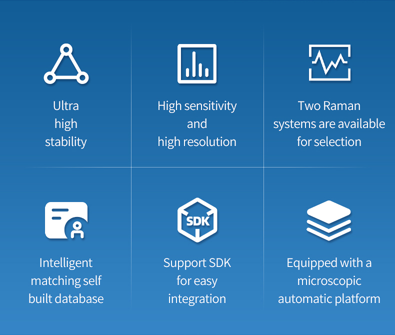
- 785 nm near-infrared excitation virtually eliminates autofluorescence from organic, biological, and colored samples — the optimal wavelength for pharmaceutical and biomedical Raman analysis
- Motorized X-Y mapping stage with 5 μm positioning accuracy and 2 μm minimum step enables automated high-resolution chemical imaging without operator intervention
- 110 mm × 75 mm travel range supports large-area mapping of pharmaceutical tablets, tissue sections, and industrial samples in a single unattended acquisition
- 1200 gl/mm grating blazed at 800 nm maximizes throughput at 785 nm excitation, delivering superior signal-to-noise performance for weak organic Raman scatterers
- Hamamatsu 11639 CMOS detector with 2048 pixels and >99.8% linearity ensures quantitative accuracy for pharmaceutical polymorph quantification and tissue biochemistry mapping
- 7 cm⁻¹ spectral resolution resolves closely related drug polymorphs (Forms I–IV), crystalline phases, and biological molecular conformations
- Integrated autofocus tracking maintains consistent focal position across uneven tablet surfaces, tissue sections, and rough industrial samples during extended mapping runs
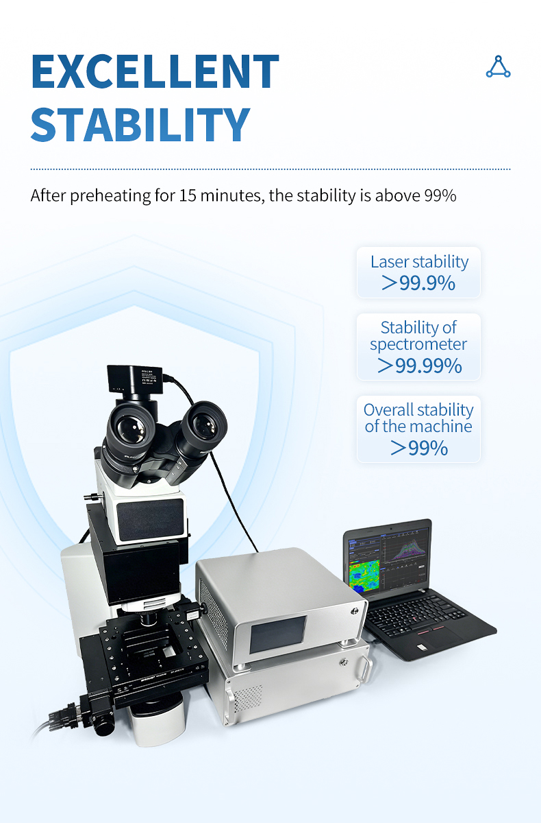
- 8-hour system stability exceeding 99% enables overnight unattended mapping acquisitions of large sample grids with reliable data reproducibility
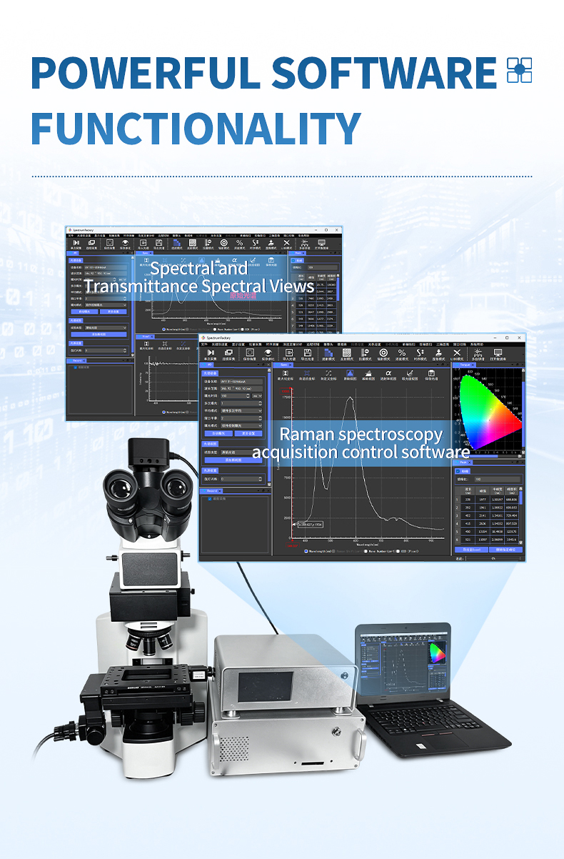
- SpectrumFactory mapping workflow — ROI selection, automated grid acquisition, spectral preprocessing (airPLS baseline, normalization), PCA clustering, and false-color chemical image export
- Rack-mounted microscope with 5x through 100x objectives and 25 μm spot size provides flexibility from survey-scale to single-cell-level analysis
- SMA905 fiber optic interface, 4 GPIO outputs, and external trigger with 5 ns latency support integration with laboratory automation and LIMS systems
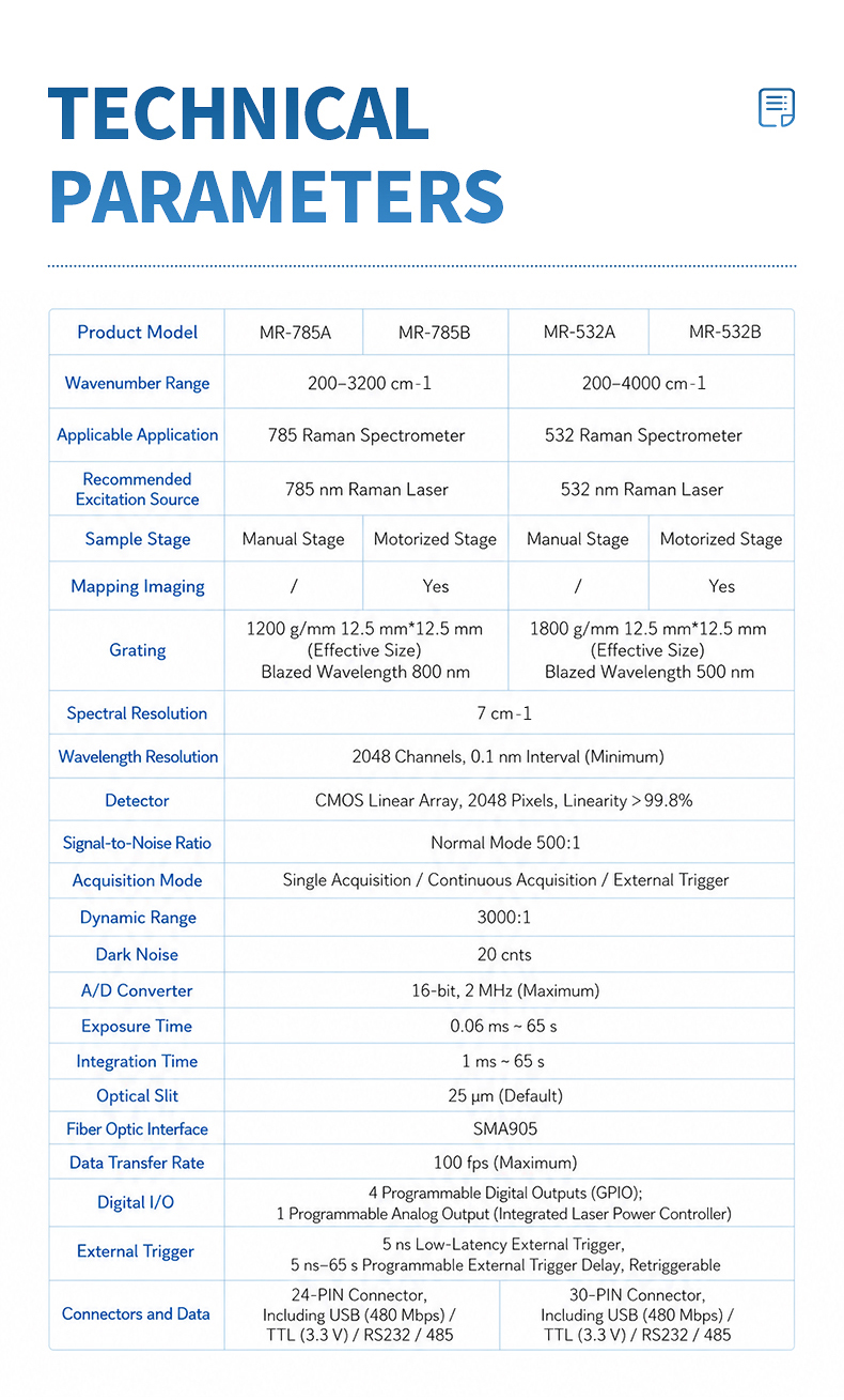
Main Parameters
| Parameter | Value |
|---|---|
| Product Model | MR-785B |
| Excitation Wavelength | 785 nm |
| Wavenumber Range | 200–3200 cm⁻¹ |
| Spectral Resolution | 7 cm⁻¹ |
| Grating | 1200 gl/mm, 12.5 mm × 12.5 mm effective size, blazed at 800 nm |
| Detector | Hamamatsu CMOS linear array, 2048 pixels, linearity > 99.8% |
| Sample Stage | Motorized mapping platform, 110 mm × 75 mm travel |
| Positioning Accuracy | 5 μm (minimum step 2 μm) |
| Microscope | Rack-mounted, 5x / 20x / 50x / 100x objectives |
| Spot Size | 25 μm |
| Reference Price | $37,000 |
Technical Parameters
| Parameter | Specification |
|---|---|
| Wavelength Resolution | 2048 channels, 0.1 nm interval (minimum) |
| Signal-to-Noise Ratio | Normal mode 500:1 |
| Acquisition Mode | Single / Continuous / External trigger / Mapping grid |
| Dynamic Range | 3000:1 |
| Dark Noise | 20 counts |
| A/D Converter | 16-bit, 2 MHz (maximum) |
| Exposure Time | 0.06 ms – 65 s |
| Integration Time | 1 ms – 65 s |
| Optical Slit | 25 μm (default) |
| Fiber Optic Interface | SMA905 |
| Data Transfer Rate | 100 fps (maximum) |
| Digital I/O | 4 programmable digital GPIO; 1 analog output (integrated laser power controller) |
| External Trigger | 5 ns low-latency, 5 ns–65 s programmable delay, retriggerable |
| Connectors | 30-pin connector, including USB (480 Mbps) / TTL (3.3 V) / RS232 / RS485 |
| Stage Travel Range | X: 110 mm, Y: 75 mm |
| Stage Positioning Accuracy | 5 μm |
| Stage Minimum Step | 2 μm |
| Autofocus | Integrated focus tracking for mapping |
| System Stability | Laser >99.9%, Spectrometer >99.99%, Overall >99% (8-hour test after 15-min warm-up) |
- Previous: MR-785A Micro Raman Spectrometer
- Next: no more
 Related Products