

 Current
Location:
HENG MEI Intelligent Manufacturing Choosing a Chemiluminescence Imaging System
Article Source: Hengmei Technology Release time:2026-11-13 13:38:00
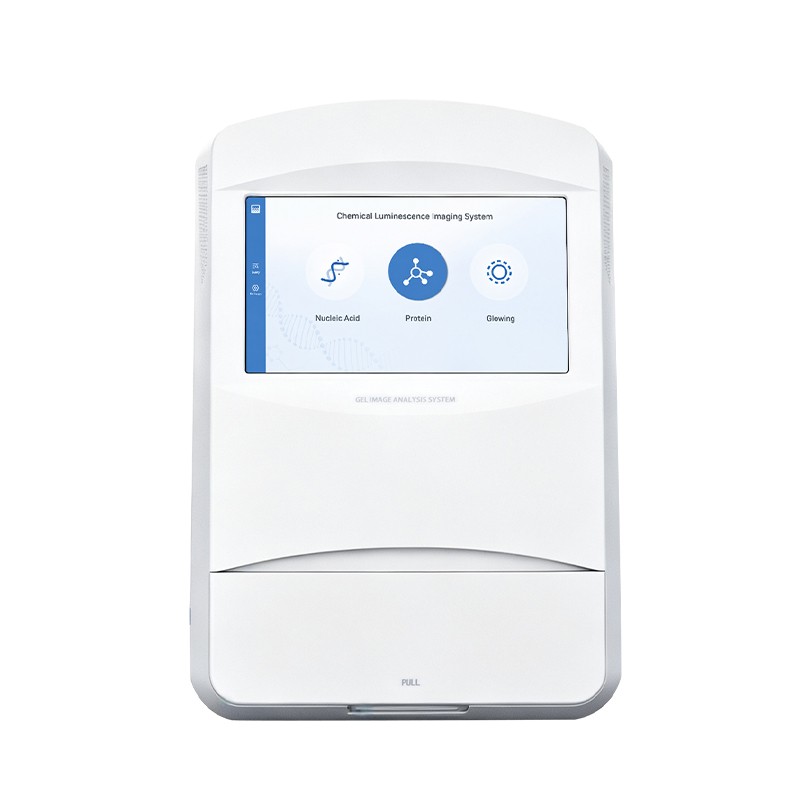
Choose a chemiluminescence imaging system by the detection chemistry, image area, camera and exposure control. HM-CL-4 combines chemiluminescence, protein gel and nucleic acid gel imaging functions. Match the imaging mode and accessories to the assay being performed.
HM-CL-4 is part of the HENG MEI Intelligent Manufacturing range supplied by Shandong Hengmei Electronic Technology Co., Ltd.
How the measurement works
HM-CL-4 captures chemiluminescent signals and provides protein and nucleic acid gel imaging modes. A cooled CMOS camera, exposure control and a 150 × 150 mm imaging field support acquisition. The selected illumination and filter arrangement must match the signal chemistry.
Choose the imaging modes used by the laboratory
List membrane and gel dimensions, detection chemistries and analysis requirements. Evaluate accessories and illumination for each mode. Plan how original images and acquisition settings will be retained and transferred. Test representative weak and strong signals to assess exposure choices, saturation handling and the analysis workflow.
Technical configuration
| Parameter | Specification |
|---|---|
| Effective imaging field | 150 × 150 mm |
| Image resolution | 3000 × 3000 pixels |
| Camera digitization | 16 bit |
Relate pixel count to the field of view
Across a 150 mm field represented by 3,000 pixels, the nominal image sampling interval is 0.050 mm per pixel, or 50 µm. This geometric calculation describes image sampling. Assess useful band detail together with optics, focus, signal and the acquisition settings for the actual gel or membrane.
Preserve the signal range needed for analysis
Inspect the acquired image for saturation and background. Use exposure settings appropriate to the signal and retain the original image before documented processing. For quantitative comparison, apply the laboratory’s normalization and analysis procedure consistently across the samples being compared.
Acquire images that support later review
Prepare the membrane or gel using the chosen detection chemistry and place it consistently within the imaging field. Review an initial exposure for signal strength, background and saturation, then select the acquisition settings required for the analysis. Preserve the original file and acquisition information before any documented processing. For comparisons across samples, keep the analysis and normalization procedure consistent and retain the basis for each adjustment. This allows a reviewer to relate the final band measurement or image presentation to the original signal captured by the instrument.
Put the selected method into practice
- Prepare the blot or gel using the corresponding detection procedure.
- Select the imaging mode and position the sample within the usable field.
- Acquire and review exposures for signal, background and saturation.
- Retain the original image and acquisition settings before performing documented analysis.
Plan support for common operating issues
| Operating issue | Checks and follow-up |
|---|---|
| Strong bands saturate | Review exposure and acquisition settings with the required analysis range. |
| Background is uneven | Review sample preparation, substrate distribution, placement and imaging mode. |
| Repeated images differ | Compare exposure, elapsed reaction time, sample position and processing settings. |
Plan the complete working configuration
Define the membrane or gel dimensions and the detection chemistries in routine use. Compare the supported illumination, filters, camera and exposure controls. Include original-image retention and analysis software in the procurement plan. Assess required accessories for each imaging mode separately when configuring a multi-application system.
Define the acceptance and reporting requirements
Agree on the following items when configuring HM-CL-4. They connect the equipment and consumables to the intended measurement and provide a basis for operator training and result review.
- Sample, detection chemistry and mode
- Exposure and camera settings
- Original image and saturation review
- Analysis version and normalization
Questions from buyers and laboratory teams
Can automatic exposure replace image review?
Review the image for saturation, background and the signal range required by the analysis.
Are gel fluorescence and chemiluminescence the same acquisition?
Select the illumination, filter and acquisition mode appropriate to the signal mechanism.
For a HM-CL-4 configuration enquiry, provide the sample type, required measurements, expected workload and reporting requirements. Include the consumables, preparation equipment and support arrangements needed at the installation site.
Address of this article:https://www.kjhm.net/news/choosing-a-chemiluminescence-imaging-system.html
 Related News
-
13 / 2026-11
HENG MEI Intelligent Manufacturing Choosing a Milk Analyzer for Collection Stations
Learn more
-
13 / 2026-11
HENG MEI Intelligent Manufacturing Choosing a Chemiluminescence Imaging System
Learn more
-
28 / 2024-04
What Can The Soil Nutrient Detector Be Measured.
Learn more
-
13 / 2026-05
HengMei SmartBuild Turbidity Meter FAQ
Learn more